38 light microscope with labels
Motility Test (Theory) - Amrita Vishwa Vidyapeetham Virtual Lab Three methods are employed for motility determination depending on the pathogenic capability of the organisms. For nonpathogens, there are two slide techniques that one might use. For pathogens, tube method can be used. I) Slide methods for non-pathogens include 1. Wet Mount slide 2. Hanging Drop slide 1. Wet Mount slide GANscan: continuous scanning microscopy using deep learning deblurring ... a A few frames from a video taken with a stage speed of 50 μm/s, with time labels indicating a slow forward movement. b Middle image of the sharp sequence a used as ground truth. c The convolution...
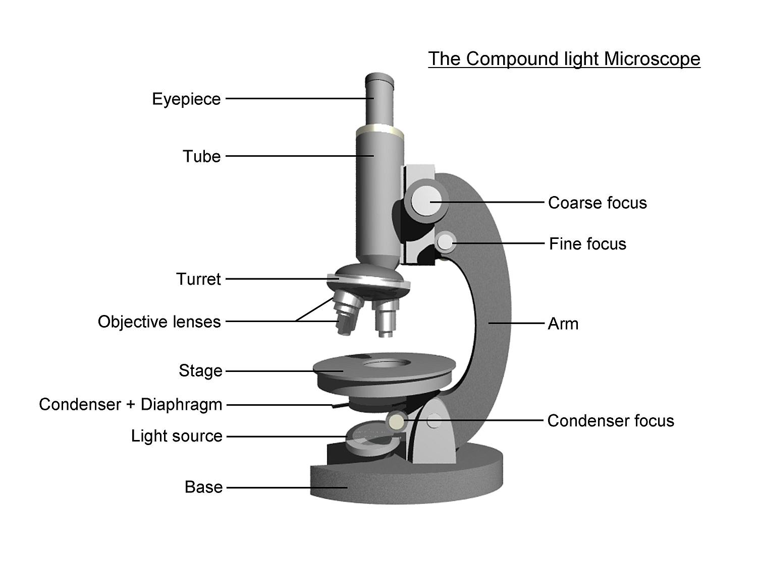
5: Get to Know the Microscope and Microbes - Biology LibreTexts To better understand the compound light microscope (brightfield), it is important to have an understanding of magnification. Commonly, compound microscopes have 4 different objective lenses: 4X. 10X. 40X. 100X. The object lens magnification is increased by the ocular lenses (located in the eyepieces), which has a 10X magnification.
Light microscope with labels
Digital Microscope Accessories 60 LED - marieclaire.fr Microscope Led Ring Light Microscope Accessories 60; B60 Series OPTIKAMICROSCOPES: LCD Digital Microscope SKYBASIC 4.3 inch 50X1000X; ... Parts of Stereo Microscope Dissecting microscope labeled; Super-Resolution Microscope System - Nikon Instruments Inc. N-SIM E is a streamlined, affordable super-resolution system that provides double the resolution of conventional light microscopes. Combining N-SIM E and a confocal microscope allows you the flexibility to select a location in the confocal image, and easily switch to view it in super-resolution, enabling the acquisition of more detail. Labeled Cell Elodea put your computer away and go to your microscope station (labeled by table number) the cell walls (which are actually double as two cells are touching) give each cell a clear outline some of the main parts of a cell include: **make a drawing of one elodea cell as you observe it under high power vanilla tweaks bedrock **make a drawing of one …
Light microscope with labels. MitchellknoeHampton The compound microscope is also termed as or called as light microscope because many lenses are present in the microsco… Cair Masakan Nak wallpaper. ... Labels 2 4 Acknowledgement Bahasa Benar Bentuk Buat Bunga Cair Cara Coklat Contoh Di Example Gaming Group in is Jadi Karangan Ke Lawatan Mandi Masakan scheme work biology - Free KCPE Past Papers Introduction to light microscope. By the end of the lesson, the learner should be able to: Define a cell; Draw and label the light microscope; Description of a cell; Drawing and labeling the light microscope . Light microscope; Diagram of light microscope; Comprehensive secondary Biology students Bk. 1 page 17; Teachers bk. 1 pages 11-19; KLB ... STED Microscopy: Turning Molecules Off And On Stimulated emission depletion microscopy (STED) is a popular super-resolution microscopy technique that researchers use to magnify microscopic organic compounds and proteins. This method is superior to conventional light microscopy as the images have less noise and are sharper given that the inspected molecule is a living organism. microscope | Types, Parts, History, Diagram, & Facts The most familiar type of microscope is the optical, or light, microscope, in which glass lenses are used to form the image. Optical microscopes can be simple, consisting of a single lens, or compound, consisting of several optical components in line. The hand magnifying glass can magnify about 3 to 20×. Single-lensed simple microscopes can ...
Super-Resolution Microscope System - Nikon Instruments Inc. The N-SIM S Super Resolution Microscope is a unique high-speed structured illumination system that achieves acquisition speeds of up to 15 fps, enabling fast biological processes to be captured at twice the spatial resolution of conventional light microscopes (up to 115nm in XY). Combining the N-SIM S and a confocal microscope gives you the ... Microscopic Morphology - BIO 2410: Microbiology - Baker College What are the names given to the groups of cells labeled 1, 2, and 3? Shape 4A A1 Diplococcus A2 Coccus A3 Staphylococcus . Shape 5 ... The microscope images in this section show different bacterial structures visible using the light microscope. All images were photographed at 1000x magification. Structure 1 1. What bacterial structure appears ... Difference Between Bright and Dark Field Microscope In a bright field microscope, the background remains bright. In a dark field microscope, the background is supposed to be dark. Specimen. Its specimen is needed to be dark. Its specimen is needed to be bright. Condition of specimen. Here the specimen should be fixed and stained. Here the specimen is not needed to be stained. White, red, and blue signals alert you to dangerous germs Introducing antibodies that specifically bind to bacteria into nanometer-scaled hybrid structures of polymer-coated metal nanoparticles and then using these structures as test labels, OMU scientists successfully detected food poisoning bacteria E. coli O26, E. coli O157, and S. aureus as white, red, and blue scattered light under the microscope.
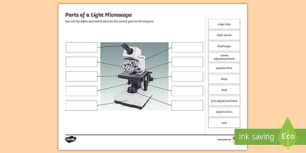
Parts of a light microscope | Teaching Resources pptx, 716.69 KB A simple worksheet suitable for low ability or SEND pupils. Appropriate for KS3 and KS4 biology lessons. Choose the parts of the micrscope from the keywords box to fill in labels pointing to parts of a microscope. 2 worksheets available - One with first letter of some of the parts already in the boxes. Answers on the final slide. Chapter 1 / Exam 1 Flashcards | Quizlet Light microscope use visible light to illuminate specimens and allow biologists to see for the first time the intricate structure that underpins all living things limited fineness of detail Electron microscope use beams of electrons instead of beams of light as the source of illumination (have shorter wavelengths) Fluorescent microscope Microscope, Microscope Parts, Labeled Diagram, and Functions Majority of high quality microscopes used in laboratory include an Abbe condenser with an iris diaphragm. When iris diaphragm is combined with Abbe condenser, it control both the quantity of light applied as well as focus on the specimen. Aperture: It is the hole in the stage through which the base (transmitted) light reaches the stage. White, red, and blue signals alert you to dan | EurekAlert! image: introducing antibodies that specifically bind to bacteria into nanometer-scaled hybrid structures of polymer-coated metal nanoparticles and then using these structures as test labels, omu...
Parts of a Light Microscope Activity | Labeling Task
IBSC at UC Santa Cruz - Instrument Details - Microscopy Leica DM5500 B Widefield Microscope. Best suited for thin (less than 10 microns) samples mounted on a standard microscope slide with a coverslip. Non-labeled, fluorescently-labeled, and colorimetric-dye-labeled samples may be used. Stage: motorized stage for 3D stack, tiling, and multi-point image acquisition.

microscope | Types, Parts, History, Diagram, & Facts | Britannica
White Light Lnterferenc Microscope Market Restraints, Segmen... MarketInsightsReports has recently launched a latest report on White Light Lnterferenc Microscope Market 2022
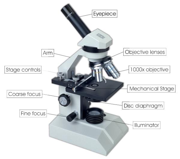
Parts of a microscope with functions and labeled diagram
White, red, and blue signals alert you to dangerous germs Using these composites as test labels bound to specific bacteria, the researchers detected food poisoning bacteria E. coli O26, E. coli O157, and S. aureus as white, red, and blue scattered light,...

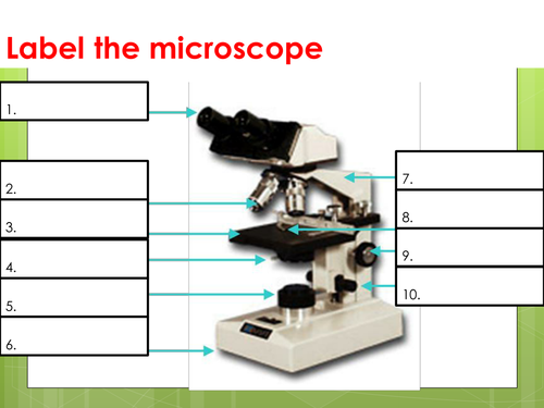
Label the microscope — Science Learning Hub
Gray Optics: Redesigning multi-channel laser scanning microscopes ... Product engineering, assembly, measurement, and testing teams handle each custom problem-solving target as an independent innovation partnership, finding the optimal solution for each challenge. Labels: Gray Optics, laser scanning, microscope, prototyping, design, microscopy Back to News Illuminating Products

Compound Microscope Parts – Labeled Diagram and their ...
Light Microscope (Theory) - Amrita Vishwa Vidyapeetham Light microscopes can be classified into Bright field microscope, Phase contrast microscope, Dark field microscope and Fluorescence microscope. Light Microscopy Light microscope uses the properties of light to produce an enlarged image. It is the simplest type of microscope. Based on the simplicity of the microscope it may be categorized into:
simple light microscope labeled - Clip Art Library
A new holographic microscope allows scientist | EurekAlert! A new holographic microscope allows scientists to see through the skull and image the brain The new label-free deep-tissue imaging with the wave correction algorithm retrieves the fine neural...

Compound Microscope- Definition, Labeled Diagram, Principle ...
How to examine histology slides: Techniques and tips | Kenhub Light microscope 2. Calibration: Place the slide under the microscope and calibrate the microscope so that the image produced is clear. Move the slide around so that its entire surface can be seen and check it with different lenses and magnifications. These objectives are usually standardized at 4X, 10X, 40X and 100X magnifications.
Parts of a Microscope Quiz
White, red, and blue signals alert you to dangerous germs! Introducing antibodies that specifically bind to bacteria into nanometer-scaled hybrid structures of polymer-coated metal nanoparticles and then using these structures as test labels, OMU...
Transmitted light microscope B3 Professional series B3-220ASC ...
A new holographic microscope allows scientists to see through the skull ... A new holographic microscope allows scientists to see through the skull and image the brain The new label-free deep-tissue imaging with the wave correction algorithm retrieves the fine neural...

Living Environment Course
A new holographic microscope allows scientist | EurekAlert! A new holographic microscope allows scientists to see through the skull and image the brain The new label-free deep-tissue imaging with the wave correction algorithm retrieves the fine neural...

Microscope Labeling Activity - SMART Board Activity - Interactive Review
High-performance 937-nm laser lets scientists see deeper with lower power The 937-nm laser design is suitable for high-sensitivity deep-tissue imaging of multiple fluorescence proteins. The laser light source provides two-photon excitations on multiple biological tissue...

Microscope Labeling #1 Diagram | Quizlet
Labeled Cell Elodea put your computer away and go to your microscope station (labeled by table number) the cell walls (which are actually double as two cells are touching) give each cell a clear outline some of the main parts of a cell include: **make a drawing of one elodea cell as you observe it under high power vanilla tweaks bedrock **make a drawing of one …
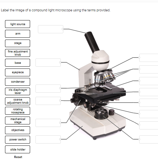
Solved PLEASE HELP THANK YOU- LIGHT SOURCE WILL BE NUMBER 1 ...
Super-Resolution Microscope System - Nikon Instruments Inc. N-SIM E is a streamlined, affordable super-resolution system that provides double the resolution of conventional light microscopes. Combining N-SIM E and a confocal microscope allows you the flexibility to select a location in the confocal image, and easily switch to view it in super-resolution, enabling the acquisition of more detail.
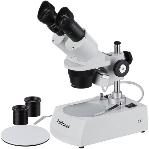
Dual Halogen Light Stereo Microscope with 20X to 80X Magnification - AmScope
Digital Microscope Accessories 60 LED - marieclaire.fr Microscope Led Ring Light Microscope Accessories 60; B60 Series OPTIKAMICROSCOPES: LCD Digital Microscope SKYBASIC 4.3 inch 50X1000X; ... Parts of Stereo Microscope Dissecting microscope labeled;

Compound Microscope Parts – Labeled Diagram and their ...

The Microscope Types of Microscopes Compound light microscope ...

Label the diagram of the microscope and explain the role of ...
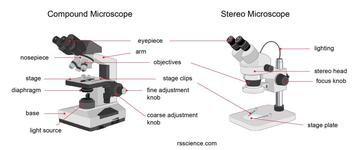
Parts of Stereo Microscope (Dissecting microscope) – labeled ...
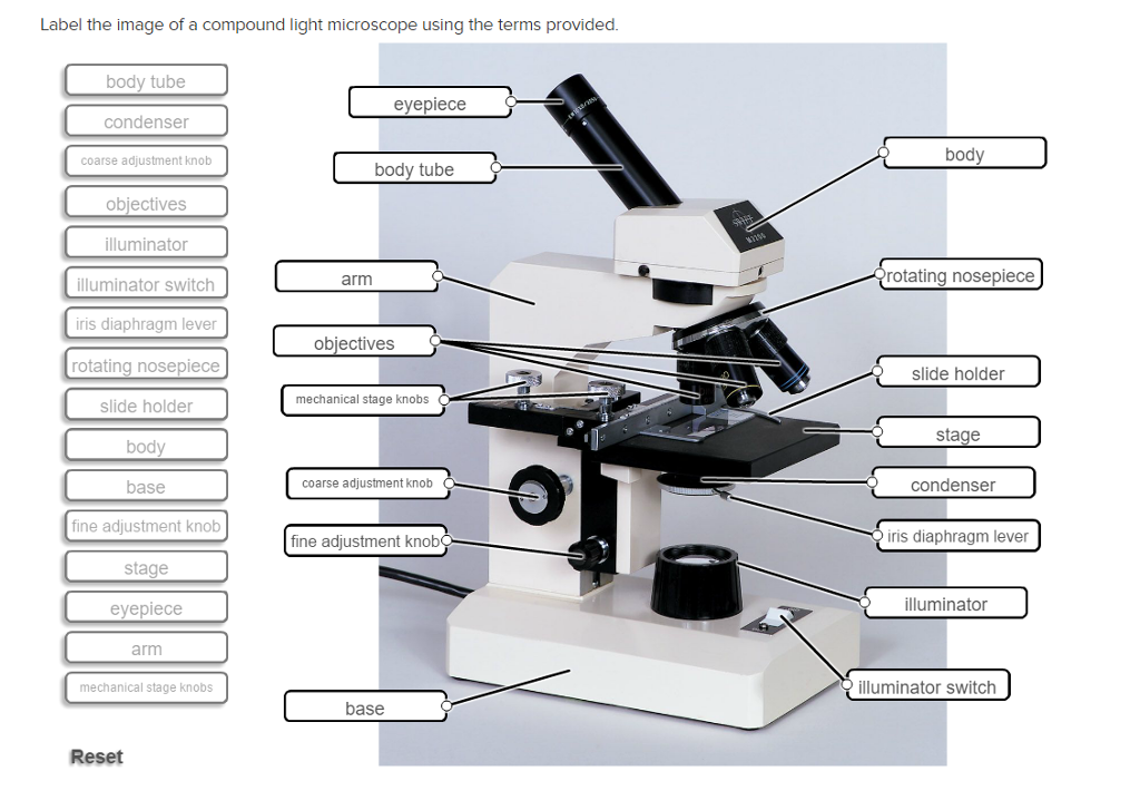
Solved Label the image of a compound light microscope using ...

Compound and Stereo- microscopes - Microscopes 4 Schools

Parts of a microscope with functions and labeled diagram
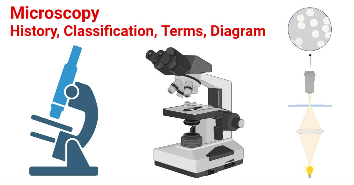
Microscopy- History, Classification, Terms, Diagram

Label Microscope Diagram - EnchantedLearning.com

Parts of a Microscope - SmartSchool Systems

National 131-LED-MS Compound Microscope 131-LED-MS B&H Photo

Microscope labeled diagram
Microscope - Teaching resources

Dissecting Stereo Microscope Parts and Functions
The Compound Light Microscope Label the following parts on ...

Parts of a Microscope Labeling Activity

Labeled Microscope Diagram | Microscope parts, Science fair ...
Microscopes: Labelling of light microscopes and difference ...
1.2: Microscopes - Biology LibreTexts

label microscope diagram | Charts | Microscope, Anatomy bones ...

Microscope labeling
Free Microscope, Download Free Microscope png images, Free ...
The microscope - Teaching resources

National Optical 40X-1000X Compound Microscope Set with Slides for Students and Kids Biology Cordless Beginner Microscope All Metal
Post a Comment for "38 light microscope with labels"